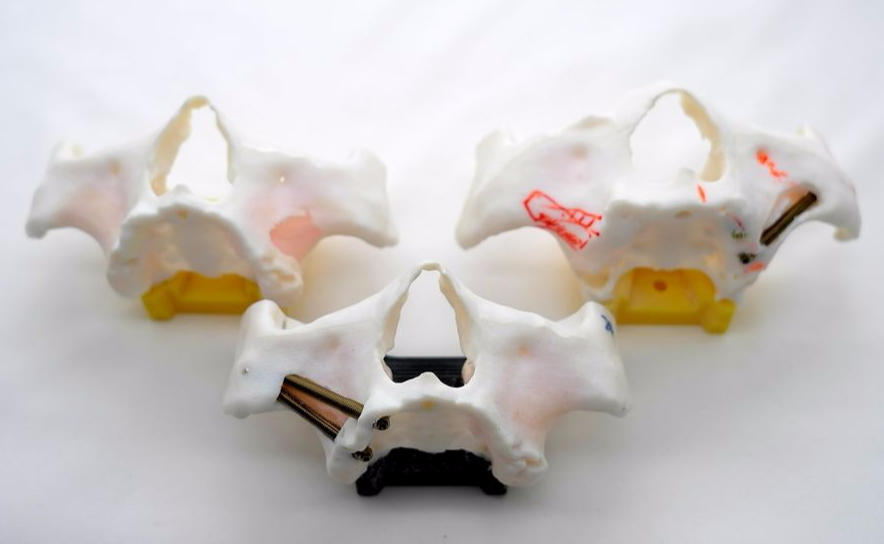
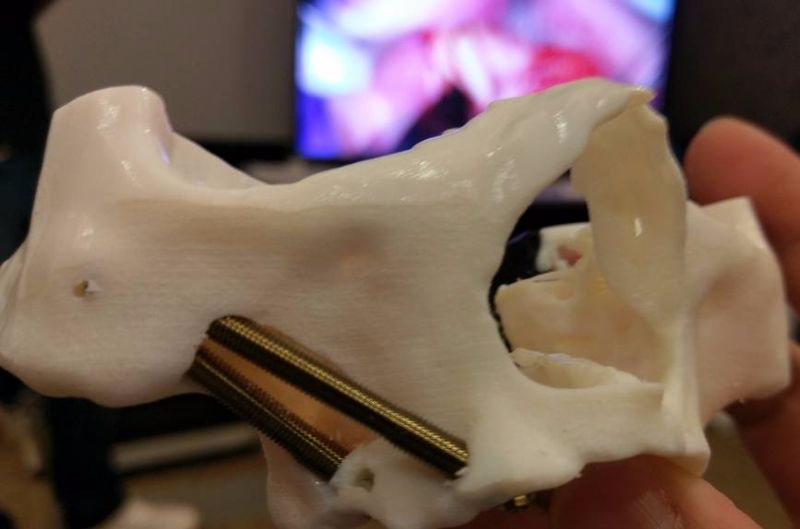
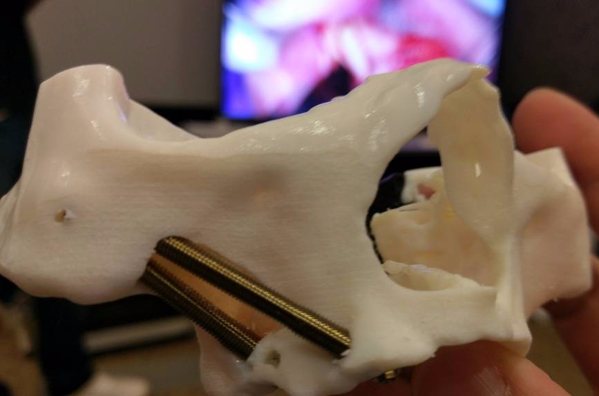
The zygoma implants are designed specifically for the upper jaw (maxilla) that has experienced severe bone loss and they make use of the cheek bone (zygoma) that sits above the upper jaw. This revolutionary dental implant technique
eliminates the need for bone grafts altogether by instead having the implants directly embedded into the cheekbone. With this procedure, patients heal much faster, and treatment time is minimal.
Improper operations
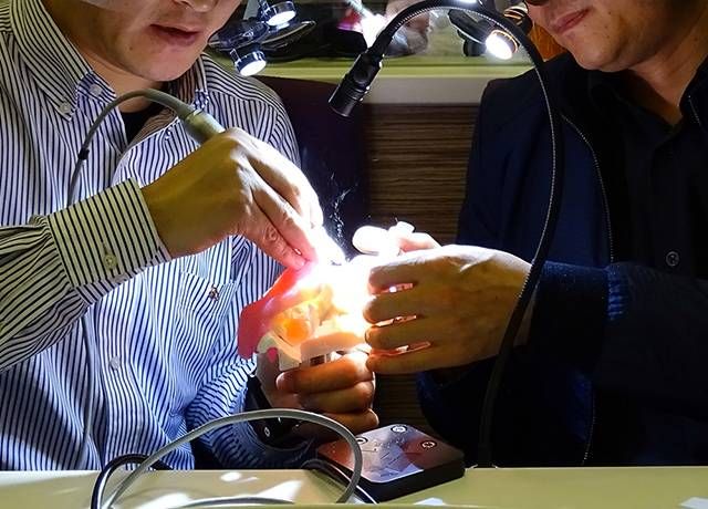
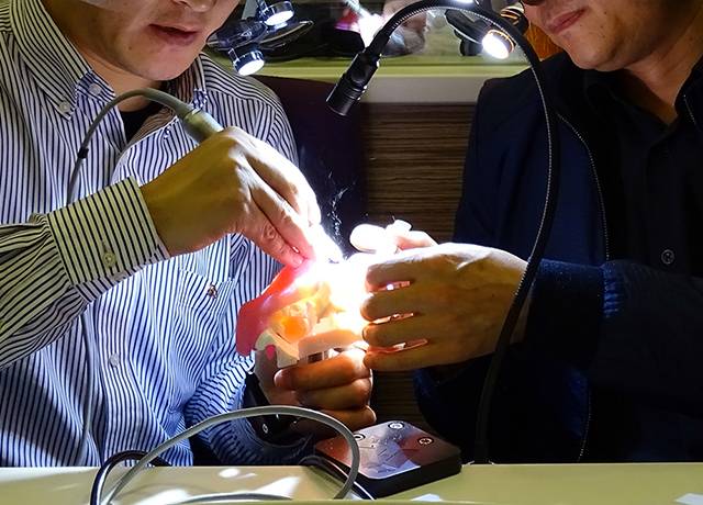
performed by a novice surgeon may lead to failure cases or serious complications. Our training model is an excellent way to learn and improve relevant skills and proficiency of dental students under experienced surgeon's instruction
without causing pain and remorse on real patient.
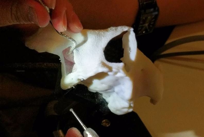
Multi-material to simulate bone & sinus properties Bone and sinus membranes at the operation zone is thin and attached to each other yet separable like real patient anatomy. The
sinus membrane is to be prevented from puncturing during the operation as in real surgery.
Practice and Review Every step of the way throughout the zygomatic implant operation on the model reveals the skillfulness and prepareness of the practicing students and surgeons at different levels. Making revision and reiteration of the procedures more qualitative so that faster improvement can be made.
CT image data of real cases Scanned patient data is converted into 3D printable format with careful design and post process to ensure best results attainable from our 3D printed training model.
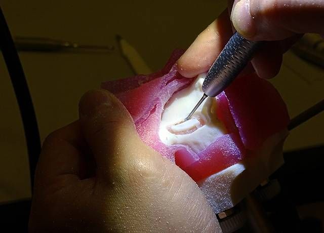
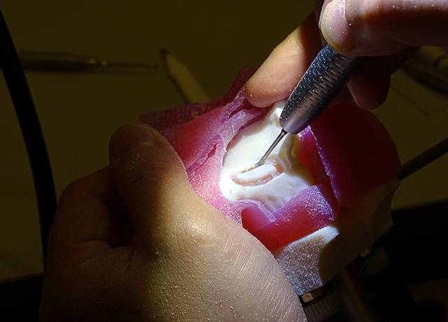
After a palatal incision of the soft tissue along the entire maxillary crest, the soft tissue is completely reflected from maxillary crest to zygomatic buttress and the suborbital nerve identified.
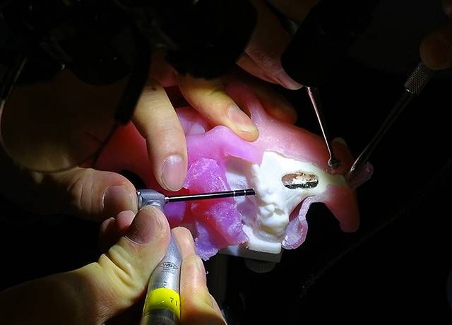
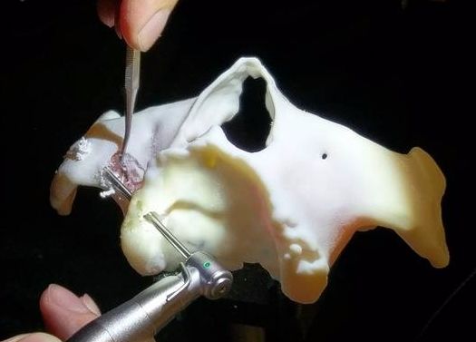
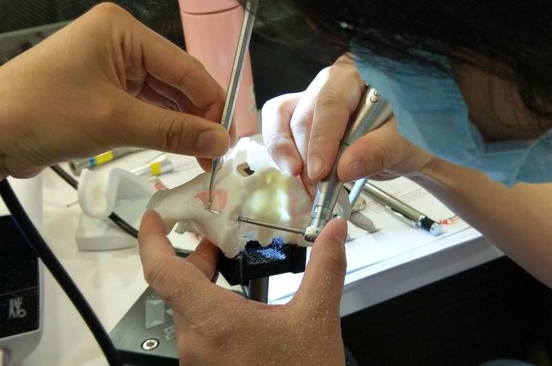
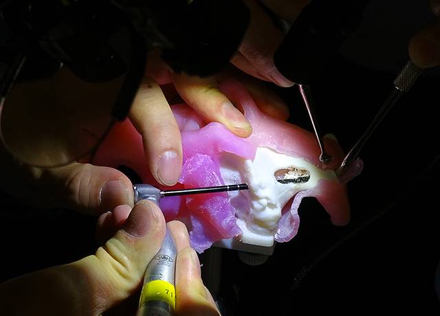
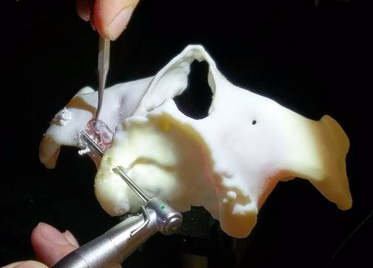
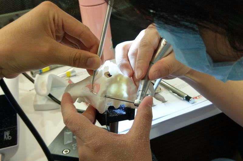
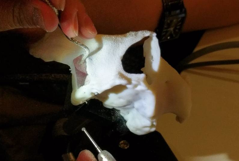
A window is then made by drilling at the upper limit between the zygoma and the sinus to determine the orientation of the zygoma and to reflect the Schneidarian membrane. The sinus mucosa is dissected from the interior sinus wall to ensure that the implant will not pass through the sinus.
To place the zygomatic implant into the prepared bone site, the drilling unit is used with a low speed level. At this stage, the angulation of implant body has to be confirmed until the apical part of implant body reaches the cortical layer of the zygoma.
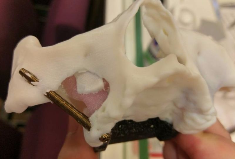
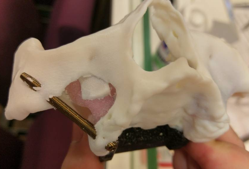
Different drills are used with increasing diameters, ending with the insertion at low speed of the self-tapping zygomatic implant..
The handle is used to rotate the implant body so that the desired depth and implant head position are achieved. After insertion of the implant, a cover screw is placed on the top of the zygomatic implant and the soft tissue closed.